- Undergraduate
Bachelor's Degrees
Bachelor of ArtsBachelor of EngineeringPartner School Dual-DegreeUndergraduate AdmissionsUndergraduate Experience
Quick Links
- Graduate
Doctoral Degrees
Doctor of PhilosophyPhD Innovation ProgramDoctor of Medicine-PhDGraduate AdmissionsGraduate Experience
Quick Links
- Research
Research by Program Area
Quick Links
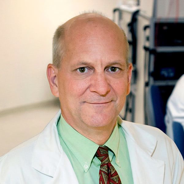
Keith D. Paulsen
MacLean Professor of Engineering
Professor of Radiology & Surgery, Geisel School of Medicine
Scientific Director, Center for Surgical Innovation, Dartmouth-Hitchcock
Co-Director, Translational Engineering in Cancer Research Program, Dartmouth Cancer Center
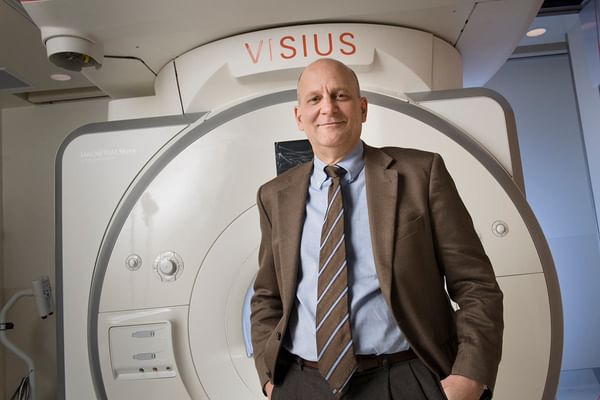
Paulsen is Scientific Director of the Center for Surgical Innovation at Dartmouth-Hitchcock Medical Center
Research Interests
Biomedical engineering; numerical methods in electromagnetics; cancer therapeutics; medical imaging methodologies; bioelectromagnetics
Education
- BSc, Biomedical Engineering, Duke University 1981
- MS, Engineering Sciences, Dartmouth College 1984
- PhD, Engineering Sciences, Dartmouth College 1986
Startups
CairnSurgical
Co-Founder
Co-Founder
Research Projects
-
Image-based registration and intraoperative updating for guiding spine surgery
Image-based registration and intraoperative updating for guiding spine surgery
This project develops and evaluates image-based registration methods involving stereovision for registration and image updating during open spine surgeries for degenerative spondylolisthesis.
-
Preoperative image updating for guidance during brain tumor resection
Preoperative image updating for guidance during brain tumor resection
The goal of this project is to optimize and validate, in human and animal studies, intraoperative image updating methods that assimilate intraoperative data acquired with stereovision and ultrasound with computational modeling to update preoperative MRI in order to maintain image registration accuracy throughout surgery.
-
Near-infrared imaging
Near-infrared imaging
Near-infrared imaging (NIR) provides a way to quantify blood and water concentrations in tissue, as well as structural and functional parameters. Since normal tissue, benign tumors, and malignant tumors each carry different concentrations of both hemoglobin and water, and have different levels of oxygen demand and ultrastructural scattering, NIR spectroscopy can be combined into standard imaging systems as an effective method of to provide additional information for breast cancer detection and diagnosis. Work is ongoing to improve techniques for better image reconstruction, display and integration with magnetic resonance imaging (MRI) and computed tomography (CT) imaging.
See also Center for Imaging Medicine
-
Magnetic resonance elastography
Magnetic resonance elastography
Magnetic resonance elastography is being developed as a technique to measure the elasticity of tissue in vivo by gently shaking the tissue in a magnetic resonance imager. The displacements measured are used to determine tissue mechanical properties which can help identify and classify breast lesions. See also Discovering at Thayer School.
-
Microwave imaging spectroscopy
Microwave imaging spectroscopy
Microwave imaging spectroscopy (also commonly referred to as microwave computed tomography) presents the challenge of measuring the signal data necessary to produce meaningful images. Development of site-specific antenna arrays along with improved electronic signal detection technology is rapidly making this measurement feasible for breast cancer detection. A second-generation tomographic breast imaging system has been completed and the data are being used to recover permittivity and conductivity maps of the breast for evaluation by a clinician.
-
Optical molecular imaging
Optical molecular imaging
Optical molecular imaging is being used to provide molecular guidance in cancer surgery. Fluorescent contrast agents are in pre-clinical and clinical studies to image cancer tumors in vivo, with a dual focus, first on getting more accurate information out of the tissue, and secondly to provide better information about the specificity of the molecules as markers. Systems and algorithms for diffuse fluorescence imaging of tissue are studied, both as a stand-alone system, and as coupled to magnetic resonance imaging and computed tomography imaging. Tracer kinetic modeling is also being developed to allow quantitative imaging of molecular binding in vivo.
-
Therapy monitoring
Therapy monitoring
Therapy monitoring is an important emerging application of imaging modalities. These and other current research topics include:
- near-infrared imaging of brain tissue;
- near-infrared spectroscopy for diagnosing peripheral vascular disease;
- electrical impedance spectroscopy for radiation therapy monitoring;
- magnetic resonance elastography for detecting brain or prostate lesions; to follow the progression of diabetic damage in the foot; and to answer basic questions of wave propagation in tissue;
- microwave imaging spectroscopy for hyperthermia therapy monitoring, brain imaging, and detection of early-stage osteoporosis;
- electrical impedance tomography for monitoring traumatic brain injury progression and therapy.
-
Fluorescence-guided surgery
Fluorescence-guided surgery
Fluorescence-guided surgery is important for the resection of some types of cancerous tumors where the tumor and normal tissue are similar in appearance and texture, and patient prognosis depends heavily on the completeness of resection. By selectively tagging tumor tissue with fluorescent dyes, it becomes possible to visually discriminate between normal and tumor tissues and improve significantly the completeness of tumor resection.
-
Non-linear image reconstruction techniques
Non-linear image reconstruction techniques
Non-linear image reconstruction techniques is at the core of the medical imaging projects. Excitation-induced measurements from each instrument are compared with calculations from corresponding numerical models to compute updated property images of the biological target. As the images are progressively updated (or refined) in a non-linear iterative process, important features and functional information related to the objects physiological status—tumor, benign tissue, etc.—become more apparent. The computational core of the breast imaging project works synergistically with all four groups to improve our fundamental understanding of these mathematical systems to improve overall image quality and resolution. These processes have been developed for both 2D and 3D geometries in each modality and are being expanded to exploit emerging parallel computing capabilities.
-
Image-guided neurosurgery
Image-guided neurosurgery
Image-guided neurosurgery gives the surgeon the ability to track instruments in reference to subsurface anatomical structures. Using clinical brain displacement data, a computational technique is being developed to model the brain deformation that typically occurs during neurosurgery. The resulting deformation predictions are then used to update the patient's preoperative magnetic resonance images seen by the surgeon during the procedure.
-
Quantitative scatter imaging
Quantitative scatter imaging
Quantitative scatter imaging makes use of the fact that normal functioning cells and cancerous cells show differences both in the components within the cells and in the structural organization of the cells within the tissue. These physical distinctions in biological structure have been shown to scatter light differently. This study develops a new approach to imaging scatter-based contrast over a wide field of view in tissue using high frequency structured light. These scattering features may provide a method to diagnostically identify abnormal tissue without the need to administer targeted compounds. This approach has the potential to generate new diagnostic screenings and new approaches for surgical guidance.
Patents
- Device and method for determining depth and concentration of a subsurface fluorescent object | 11,662,315
- Method and apparatus for quantitative hyperspectral fluorescence and reflectance imaging for surgical guidance | 11,656,448
- Specimen imaging with x-ray and optical measurement | 11,633,145
- Method and apparatus for medical imaging using differencing of multiple fluorophores | 11,564,639
- Method and apparatus for quantitative and depth resolved hyperspectral fluorescence and reflectance imaging for surgical guidance | 11,510,600
- Hand-held stereovision system for image updating in surgery | 11,406,471
- Systems and methods for guiding tissue resection | 11,395,704
- Systems and methods for guiding tissue resection | 10,667,870
- Systems and methods for cardiovascular-dynamics correlated imaging | 10,575,792
- Surgical navigation with stereovision and associated methods | 10,568,535
- Apparatus and methods for structured light scatteroscopy | 10,485,425
- Structured-light imaging systems and methods for determining sub-diffuse scattering parameters | 10,463,256
- Method and apparatus for calibration of stereo-optical three-dimensional surface-mapping system | 9,456,200
- Method and apparatus for determining tumor shift during surgery using a stereo-optical three-dimensional surface-mapping system | 9,336,592
- System and method of for providing patient registration without fiducials | 9,179,888
- System and method for registering ultrasound and magnetic resonance images | 9,098,904
Courses
- ENGG 325: Introduction to Surgical Innovation
- ENGM 189.1: Medical Device Commercialization (.5 credit)
- ENGM 189.2: Medical Device Development (.5 credit)
- ENGS 105: Computational Methods for Partial Differential Equations I
- ENGS 205: Computational Methods for Partial Differential Equations II
Books


Videos
Seminar: Dartmouth's Center for Surgical Innovation
IMRIS Dartmouth iMRI & iCT
Breast Cancer Imaging Techniques
Graduate Student Engineering Research: Breast Cancer Screening
Graduate Student Engineering Research: Diagnosing Hydrocephalus
Seminar: From Biomedical Computation to Imaging and Back Again: Lessons Learned and Advances Gained from Two Decades of Research at Thayer
News


In the News
NH Business Review
Dartmouth Cancer Researchers' Cancer Surgery Innovation Named Product of Year
Nov 04, 2021
Dartmouth Cancer Researchers' Cancer Surgery Innovation Named Product of Year
Nov 04, 2021
ScienceDaily
Diagnostic screening: Microwave imaging of the breast may be better and safer
Dec 16, 2014
Diagnostic screening: Microwave imaging of the breast may be better and safer
Dec 16, 2014